The Legacy family deeply mourns the loss of one of our founding fathers for which our name was created to honor — Ronald N. Williams, MD. October 2, 1942 – October 28, 2017
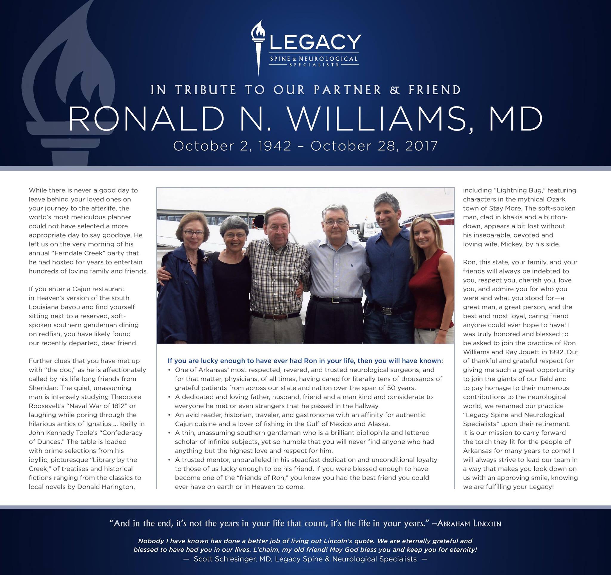
The Legacy family deeply mourns the loss of one of our founding fathers for which our name was created to honor — Ronald N. Williams, MD. October 2, 1942 – October 28, 2017
Retrospective Review of an MIS Lumbar Fusion Technique in Outpatient and Hospital Settings
Scott Schlesinger, M.D. (Legacy Neurosurgery and Spine, Little Rock, AR)
Introduction: Advances in MIS surgery have helped expand the number of procedures that may be performed safely on an outpatient basis. Patients may benefit from decreased tissue disruption that potentially minimizes pain, blood loss, complications, hospital stay, and recovery time. Our clinic has combined MIS techniques with image guided navigation to develop a lumbar fusion procedure that can be performed through a single midline incision with cortical screw trajectory, achieving direct decompression, fusion, and instrumentation, permitting patients with low comorbidities to be treated on an outpatient basis. Here we compare surgical results from outpatient and hospital settings.
Methods: Under IRB approval, a retrospective chart review of a consecutive series of adults with lumbar stenosis and instability, treated over an 11-month period by one surgeon, was performed. Charts were reviewed for demographics, diagnosis, medical history, surgical parameters, and complications.
Results: A total of 73 patients with degenerative disease positive for radicular pain and segmental instability were reviewed (Table 1). Outpatients (n=40) were single level procedures, while hospital patients (n=33) were treated at one (n=28) or two levels (n=5). Average OR time was 204±51 minutes for outpatients and 221±61 for hospital patients (Table 2). Average estimated blood loss was 345cc±198 for outpatients and 298cc±217 for hospital patients. All surgical incisions were <2cm and no blood transfusions were required. All outpatients were successfully discharged on the same day. Average stay for hospital patients was 2.5±2.6 days. There were no unanticipated complications or trends observed.
Conclusion: Surgical outcomes in this series demonstrate the initial benefits of the midline technique in outpatient or hospital settings. Navigation, cortical bone trajectory, and keyhole incision allows direct decompression and instrumentation, optimizing MIS in the lumbar spine. Long-term, prospective clinical studies may further demonstrate the safety and long-term efficacy of this technique.
Table 1. Patient Demographics
| Outpatient (n=40) | Hospital
(n=33) |
Total
(n=73) |
|
| Age | 52.1 ± 8.6 | 62.4 ± 17.0 | 56.8 ± 14.0 |
| Sex | 24 Males
16 Females |
15 Males
18 Females |
39 Males
34 Females |
| Prior Surgery (any lumbar level) | 15 Yes (38%)
25 No |
15 Yes (45%)
18 No |
30 Yes (41%)
43 No |
| Levels Treated | All single levels | 28 single level
5 two-level |
68 single level
5 two-level |
Table 2. Surgical Parameters and Intra-Operative Complications
| Outpatient (n=40) | Hospital (n=33) | Total (n=73) | |
| Surgical Parameters | |||
| · OR Time (Minutes) | 204 ± 51 | 221 ± 61 | 212 ± 56 |
| · Est. Blood Loss (CC) | 345 ± 198 | 298 ± 217 | 324 ± 206 |
| · Hospital Stay (days) | Same Day | 2.5 ± 2.6 | 1.1 ± 2.1 |
| Intra-Op Complications (n) | |||
| · Intra-Op Durotomy | 2 | 3 | 5 |
| · Dural Fistula | 1 | 0 | 1 |
| · Cardiac Issue | 0 | 1 | 1 |
| · Urinary Stasis | 1 | 1 | 2 |
| · Psuedomemingocele Repair
(From prior fusion surgery) |
0 | 1 | 1 |
Submitted to the 2017 Annual Meeting of the Congress of Neurological Surgeons, to be held October 7-11, 2017 (Boston, MA)