Pavilion MRI
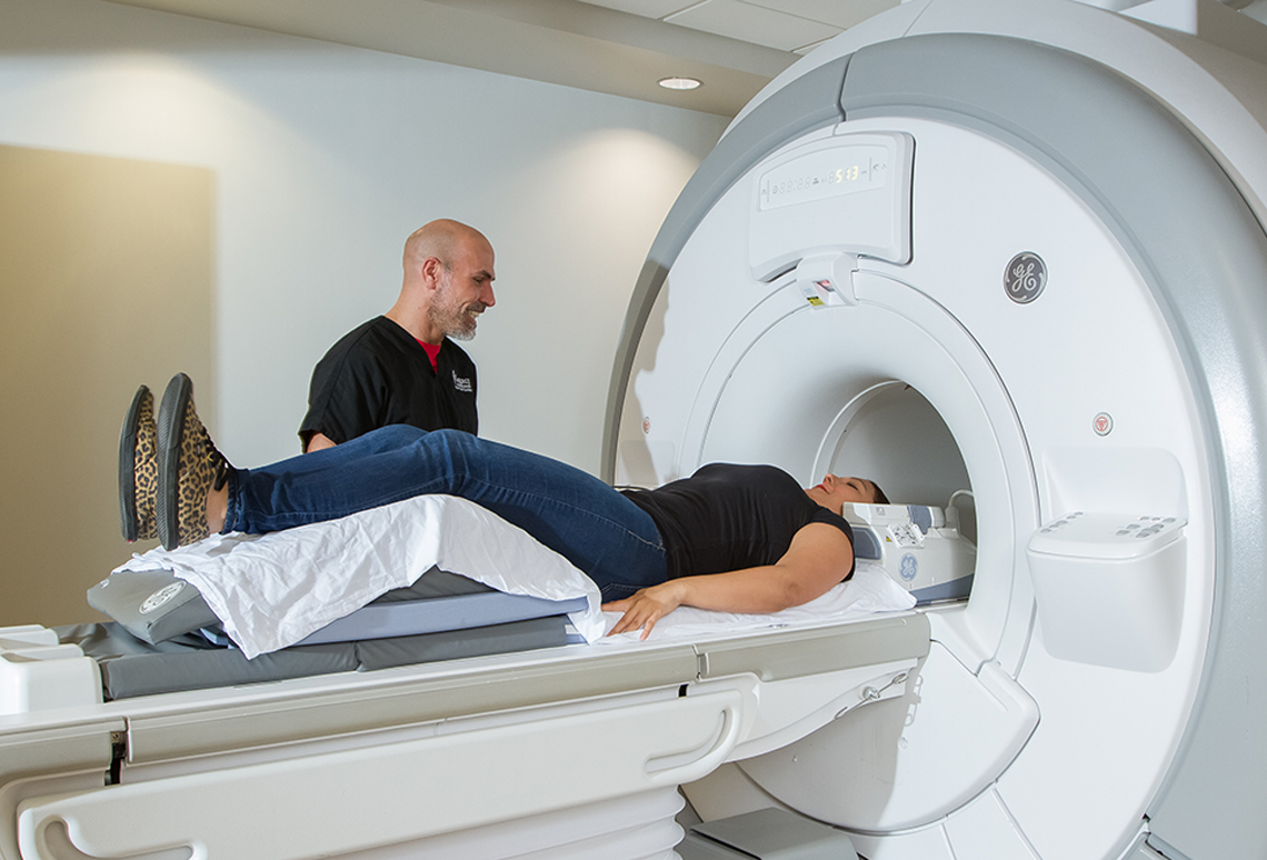
Pavilion MRI combines the latest technology in neuro-imaging with experienced, friendly technicians, physicians and staff, to create the most high-quality, comfortable MRI experience. Our clinic features a GE 1.5T “closed” MRI machine, the largest wide bore high-resolution magnet on the market. This state-of-the-art MRI is designed with a more spacious setting to help reduce patient anxiety, and is ideal for those who may be claustrophobic. The design can also accommodate larger body types (weighing up to 500 pounds). We seamlessly merge high-resolution with comfort, and provide the highest-quality images available for any area of the body, excluding the heart and breasts.
Preliminary results are available to the patient’s physician within an hour of the MRl appointment. Preliminary reports are available within minutes by a national radiologist group and sub-specialists in all body areas and systems scanned by our MRI. Experienced technicians and an on-site physician are available to ensure quality of the scanning experience and to provide sedation when needed for the claustrophobic patient.
Pavilion MRI is conveniently located in the quiet, serene and beautiful setting of the Pavilion in the Park center in Little Rock. Often, same day appointments are possible. Friendly, cooperative staff is available to assist in all aspects of scheduling and authorization.
The MRI Explained
Magnetic Resonance Imaging (MRI) is a scan used for a medical imaging procedure. It is based on differences in the physical and natural magnetic properties of protons in water molecules in different tissues in the body.
It uses a magnetic field and radio waves to take pictures inside the body. It is especially helpful to collect pictures of soft tissue such as nerves and muscles that do not show up on x-ray examinations.
The MRI scan consists of a table that slides into a large cylinder. Inside the cylinder is a powerful magnetic field.
Soft tissue contains water molecules and the magnetic field acts upon microscopic substances (called protons) found in water. The magnetized protons in the soft tissue send out an echo in response to the MRI scan’s radio waves. A computer then organizes these echoes into images.